In 1999 or 2000 I wondered what these old watches would look like if imaged by electron microscopy. I contacted the labs mentioned below, and they graciously agreed to help. One watch was sent to each lab for analysis. By clicking on the small picture, the full-size image will appear.
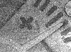
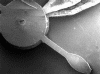
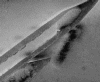
Picture #3, dial side, center post, hands.
Electron microscopy is unlike ordinary light microscopy, or photography. Essentially Scanning Electron Microscopy (SEM) involves passing some current through a thin metal filament, much like a light bulb or x-ray tube. Electrons will build up around the filament, but they won't know where to go. A charge separation is then applied, usually in the range of 0.5 to about 50 kilovolts. This causes the gathering mass of electrons to move toward the positive (anode) side of the charge separation in an accelerated fashion in the form of an 'electron current.' This causes the specimen to be bombarded with a very focused beam of high-energy electrons, while it sits in a vacuum chamber (which rules out scanning, for example, yourself).
The specimen will absorb some of the energy and will exist for some ultra-short time in an excited state. During its relaxation to ground state, it must give back some of that energy, and it does so in the form of 'flourescent electrons.' It's sort of like an electron echo. These electrons then interact with a scintillating material, which gives off light in the visible spectrum. This scintillating event is amplified by a photomultiplier system to increase brightness to the visible level, and the event is recorded.
Many of these events occur in the scan process, and eventually an image is created. The type of signal generated by the sample will depend on its constitution, as well as scan parameters, for example the kilovoltage current. Another type of electron microscopy is called Transmission Electron Microscopy (TEM), but this uses super-thin slices of sample. The electron beam goes through the specimen, rather than echoing back at you.
I am most grateful to those who helped make these images and this page possible: Doreen Ah Tye, of the National Center for Electron Microscopy, and Ken Streib and Przemyslaw Mitan, of the Materials Engineering department at Arizona State University. They enthusiastically responded to my unusual request to image these watches with their machines, and created the images you see here. Two vintage watches were sacrificed for the project.
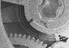
Picture #1, hand, 20x
Picture #2, hand, 135x
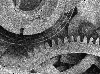
Picture #3, luminous granules, 550x
Picture #4, luminous granules, 1200x